Life-Health Sciences Internship Program
The LHSI is a program at Indiana University, Indianapolis that matches qualified students to year long internship sites to help them learn, work, and gain experiences related to their major and career aspirations.
Below is a documentation of my 2024-2025 internship and research showcase with Dr. Schlecht and lists all my progress and learning experiences as part of this program.

About My Internship Site
I received the opportunity to work with Dr. Stephen Schlecht in the Orthopedic Surgery Department at IU Indianapolis. My internship site researches sex differences specifically females’ susceptibility to ACL injuries compared to males using in vivo mice models. We use mice in our research to look at the responses to loading and injury on the ACL in terms of submaximal loading on the ligament.
We compare the results in the male and female mice and look at sex hormones, fibrotic responses, anabolic and catabolic responses, age differences and even osteoarthritis post ACL tear. Studies ranged from arthrofibrosis differences, role of gene expression, diet, synovial studies, cadaver studies, and more.
We are also engaged in ongoing collaborations with Michigan State University, Penn State, and the University of Alabama. At Michigan State University, researchers focus on human cadaveric models to better understand ACL structure and injury mechanisms. Penn State conducts work similar to our lab, investigating sex-based differences in ACL injuries, but uses in vitro ACL cell cultures, while our lab conducts in vivo studies. Meanwhile, the University of Alabama examines ACL injuries from a chemical perspective, identifying the molecular processes involved.
LHSI 2024-2025 Showcase
My Project & Abstract
IU Indianapolis, Campus Center
Sex Differences in Fibrotic Responses Following ACL Injury in Mice
This pilot study investigated sex differences in post injury response that focuses on aggressive fibrotic responses in females following an anterior cruciate ligament (ACL) tear. Using the C57BL/6J mouse strain, we examined 5 male and 5 female mice, aged 18 weeks, with ACL rupture on the right knee with the left knee serving as an internal control. 3 mice per sex were euthanized at 3 weeks post injury. The remaining 2 mice per sex were euthanized 6 weeks post injury. Our primary goal was to explore gene expression changes in response to the injury, with a particular emphasis on identifying key pathways that drive inflammation and fibrosis. Early findings from a previous study with young females indicated an aggressive fibrotic response, which limited range of motion and severely impacted recovery. Clinically, females are more prone to arthrofibrosis post ACL surgery than their male counterparts. Through this study, we sought to uncover underlying genetic and molecular factors that contributed to arthrofibrosis development and guide future treatments. This research was directly tied to a forthcoming clinical study in patients, providing insight into potential therapeutic targets for managing arthrofibrosis and improving post-injury recovery outcomes, particularly in females
Milestones: Challenges & Successes
2024 Fall Semester
"Learning the Anatomy"
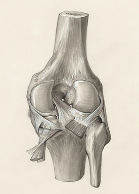
In the initial weeks of my internship, Dr. Schlecht taught me knee anatomy and how to read ACL histological images, emphasizing, "The ACL runs from the back of the femur to the front of the tibia." Recognizing this structure is crucial for effective imaging.
He also helped identify knee structures like the fat pad, patellar ligament, and other anatomical markers. He described the fat pad as "a triangle with distinct bubble features" and the patellar ligament "runs in front of both the patella and fat pad.” He also noted that the tibia is flatter and wider while the femur is rounder.
I've gained extensive knowledge about the knee's anatomy and its mechanics. Dr. Schlecht demonstrated a knee model, illustrating how the bones slide and shift as we bend our knees. This visual aid has deepened my understanding significantly and has influenced how I approach studying for my kinesiology courses. Grasping the mechanics of these structures has enabled me to apply this knowledge to my daily life.

"Imaging Histological Slides"
This semester I also learned how to use and manage our advanced microscope for histology imaging. I successfully learned operate the Nikon H550L, a high-tech motor microscope that features fluorescent lighting, computer connectivity for improved visualization and an integrated camera for capturing intricate images of tissue histology. This experience has been a key milestone, allowing me to analyze samples more effectively and contribute to our research on ACL injury susceptibility with greater accuracy. Seeking guidance from my supervisors and co-interns was invaluable; their explanations of the microscope's functions greatly facilitated my successful operation of the equipment.

2025 Spring Semester
"Making Histological Slides"
In the following semester, my supervisor assigned me a new task that required me to learn and master a new skill I began the process of slicing ACL samples to prepare them for histological slides. This task was quite meticulous, as we needed to slice the hind limbs of mice to obtain samples of the ruptured ACL, cutting them to a delicate thickness of 5-10 microns, which made them prone to breaking during transfer to the water bath. However, with consistent practice, developed a rhythm and successfully sliced the ACLs. Additionally, I used the every 30 slices to ensure I had accurately reached the ACL in the knee, making my understanding of anatomy essential. After a few days, I became both efficient and precise in my technique.

"Data Collection, Cropping, & Thresholding"
One of the main challenges I faced during my internship was adapting to technology and computer-based tasks, as I’ve never considered myself very tech-savvy. Initially, learning to use new programs like the FIJI application for cropping and analyzing histological images was quite frustrating. At one point, I cropped too much of the bone while thresholding the ACL area, which distorted the results and added to my frustration. Fortunately, my supervisor guided me in understanding how the ACL blends into the bone and shared techniques to better differentiate the tendon from the bone in that transition area. With practice and support, I became more confident in using the software and improving my analysis skills. This was an important skill to perfect because it was important to collect accurate data when thresholding the darkest staining to determine what percent of the ACL had damage in order to compare females and males injury response.
